CT Imaging

Computed tomography (CT) imaging is an x-ray imaging modality that operates by obtaining numerous two-dimensional x-ray images (projections) from all angles around a subject. A three-dimensional image of the subject is reconstructed from these projections. Clinically, CT is used for the purpose of disease diagnosis or radiation treatment planning. Additionally, there are a number of other preclinical and clinical CT modalities.
Photon-counting CT (PCCT)
Photon-counting CT (PCCT), or spectral CT, utilizes high-flux photon-counting detectors to discern the energy of x-rays incident on the detector (which is not possible with current technology). This enables a number of exciting possibilities such molecular imaging of high atomic number (Z) contrast agents, such as gold nanoparticles, simultaneous imaging of multiple high-Z contrast agents, and performance enhancements over conventional CT like higher contrast-to-noise, improved spatial resolution, and reduced radiation dose, among others.
X-ray fluorescence CT (XFCT)
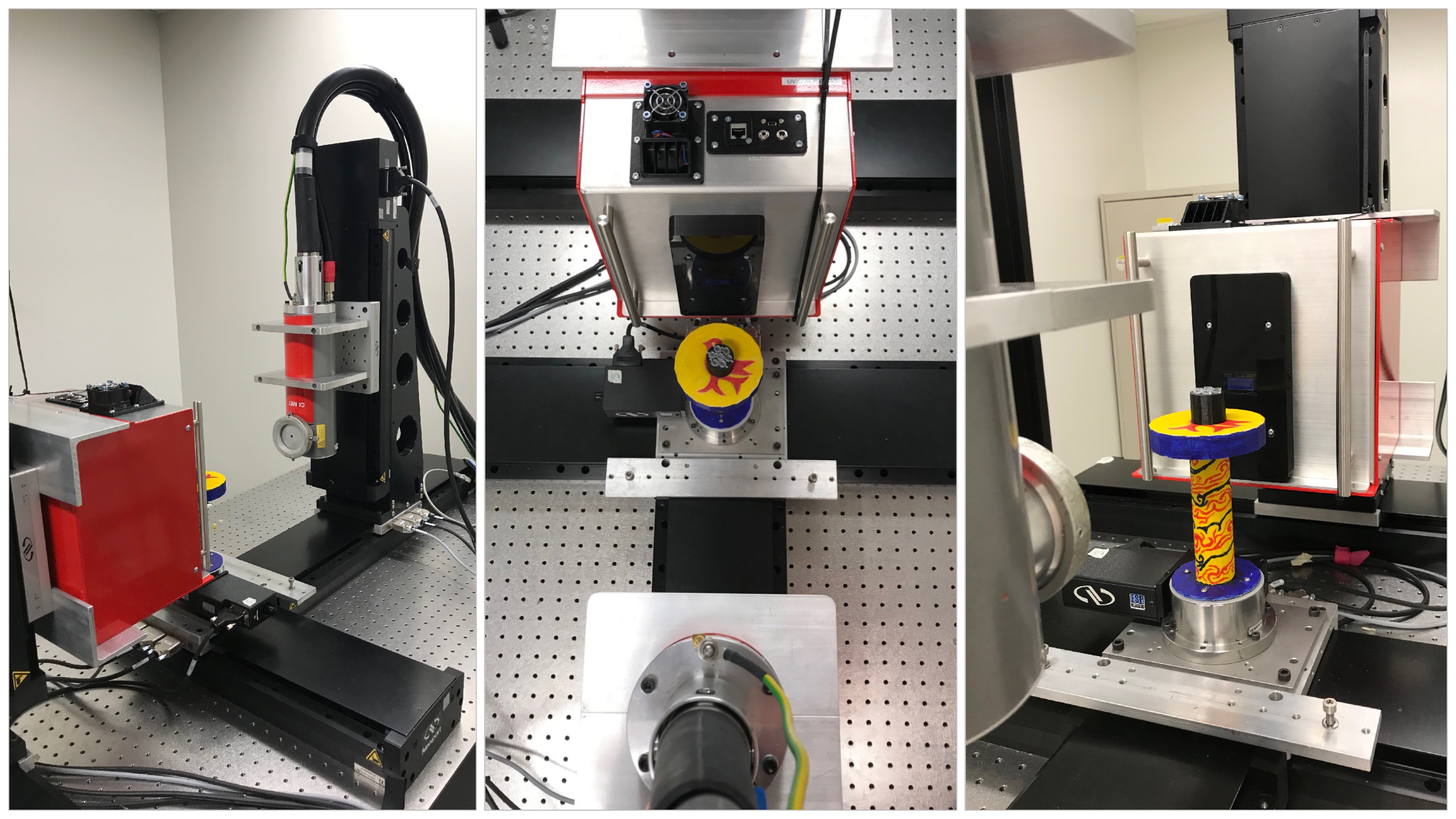
X-ray fluorescence CT (XFCT) detects the scattered x-ray beam from a subject, primarily the fluorescent x-rays generated from high-Z materials via the photoelectric effect. The photon-counting detectors used for XFCT have a high energy resolution, which allows for the visualization of high-Z contrast agents at very low concentrations. It is primarily a small animal modality.
Megavoltage cone-beam CT (MV CBCT)
Megavoltage cone-beam CT (MV CBCT) is a clinical modality primarily used in radiation therapy. It is mainly used to verify patient setup by comparing the MV CBCT image taken immediately before treatment to the CT image dataset the treatment was planned on. Current research is focused on new detector materials that could enable higher image quality at lower imaging doses.
People
Magdalena Bazalova-Carter
